
Searching for signatures in biology
The Scientific Computing Group at the Pontifical Catholic University of Goias focuses on using computational intelligence to develop solutions to biological problems.
By Rute Costa
In the middle of Brazil, away from the spotlights of São Paulo and Rio de Janeiro, is a group of scientists making groundbreaking discoveries.
The Scientific Computing Group (GCC) has been running since 2003 at the Pontifical Catholic University of Goias (PUC Goias). Their focus is on using computational intelligence to develop solutions to biological problems. They work with spectral imaging (using hyperspectral and multispectral cameras) to analyse patterns in biological materials: anything from plants to cancerous cells.
Spectral cameras can capture the complete spectrum at every point in an image. They unveil several bands of colours, as seen in a rainbow, which are produced by separation of the components of light. Whilst multispectral cameras usually only capture 3-10 bands, hyperspectral cameras are super high-definition, capturing hundreds of narrower bands.
The information from the spectral cameras gives a unique ID to each type of biological material. This research group collected numerous samples and built a computational model that is capable of analysing and recognising these patterns, known as spectral signatures.

Cancer diagnoses in a snap
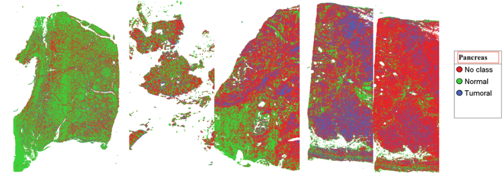
Being able to identify the spectral signature of materials opens the doors to massive improvements in medical diagnostics and research. The Research Group is pioneering the application of this methodology to the diagnosis of cancer (uterine, pancreatic and skin cancer). The current process of diagnosis is lengthy and relies heavily on the human eye to analyse microscopical images. Pathologists take weeks to diagnose cancer because they must decompose the sample and conduct a series of tests until they are able to reach a conclusion. Through spectral imaging, by contrast, the results are achieved in just a couple of hours: thirty minutes to acquire the sample, one hour and thirty minutes for the algorithm to analyse it, and it’s done. Alongside this, the scientists are developing methods to enable the diagnosis of cancer in its early stages.
From ten days to two hours
A similar model is being used to advance the identification of multiresistant bacteria, and to find out what kind of antibiotics can fight them. The most widely used process involves growing enough culture to be able to identify the bacteria and test different types of antibiotics. It takes, on average, 10 days. However, by the time patients with serious bacterial infection get to hospital, they don’t often make it past the hour. A method that takes weeks to understand what can save them is fruitless at critical times. That is why this group of scientists is developing an alternative: through hyperspectral imaging, they are pulling together a database of multiresistant bacteria and the antibiotics they are immune to. The model in development can determine the bacteria’s genome, and rule out inefficient antibiotics, in under two hours. It can save lives.
An app for an eye
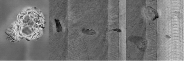
They are also developing a software to help count bacteria and speed up laboratorial procedures in this field. At the moment, cells are counted by eye – a time-consuming and unexciting task for any scientist. Hundreds of new cells every day, and numerous pairs of tired eyes, are causing severe delay in the advancement of medical science. The group is in the process of developing a simple mobile app that, from a single image, is capable of recognising different types of bacteria, of classifying and counting them, quickly and with precision. This is a huge step towards understanding and finding treatment against multiresistant bacteria.
Updating treatments
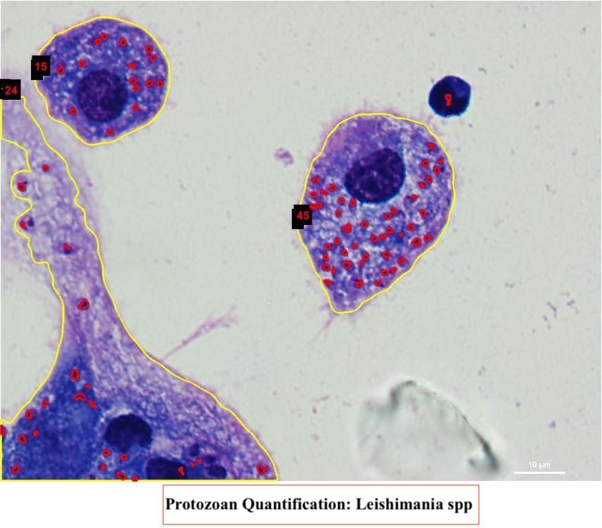
Another app currently in development gives attention to a disease that has been neglected by the pharmaceutical industry for decades: leishmaniasis, a tropical and subtropical illness caused by parasites and transmitted by the bite of sandflies. Because most of the cases of this disease occur in remote areas such as the Amazon, treatment is outdated (read: one painful injection a day for twenty days, with a 50% chance of success) and often unavailable. The research group is working with partner labs to help them speed up the process of developing a new medication. They are developing a software that can identify cases of the disease through the analysis of the microscopical image of a sample. The software can also count the protozoa (parasites that cause the illness), speeding up all lab procedures. Being mobile, the app would also allow research and treatment to occur in remote places with little to no resources. It’s revolutionary.
Their projects do not end here. The group is using methodology based on analysis and processing of spectral images to identify potential medical uses of popular plants in Brazil, to control the quality of pharmaceutical drugs, and to help identify and classify inflammatory processes. Check out their page to access the numerous projects they are currently working on.
“Alone we can’t get anywhere”
This group’s contribution to the advancement of medical research is invaluable, and they want to open it to the world. Although they have links to the major research labs in Brazil, the group still finds itself isolated from the global picture. Arlindo Galvão, one of the scientists, reveals their working motto: “alone we can’t get anywhere”. They are looking to share their knowledge internationally and let it be challenged, eager to work collaboratively to find solutions to the medical problems affecting humanity. Join them.
Photo credit: The Scientific Computing Group